|
|
|
|
|
|
|
Muscle Spindle Sensory Receptors
|
 |
Location: Muscle spindles are distributed throughout skeletal muscle bellies, attached to the outside of the perimysial wall surrounding fascicles; so they are actually in the corridors between fascicles, along with lymph ducts, nerves, capillaries, and other structures. This way, the spindle monitors the contraction of the fascicle, and is getting an average of all the cells within. In other words it samples a whole group of muscle fibers. We could say it operates on hearsay, without a direct witness. The spindle controls and reports on the extrafusal muscle fibers inside their fascicle, based on what the fascicle fascia says is going on inside. It’s like someone monitoring scuttlebutt on the other side of a wall.
Why couldn’t they have been placed inside the fascicles, right next to the individual fibers, attached to endomysium in order to offer first person witness? Because there’s no room for them in there; too much goes on inside the fascicle. Nevertheless the spindles do manage to take proper information from the perimysial wall; the malfunction occurs in the relay system to the brain.
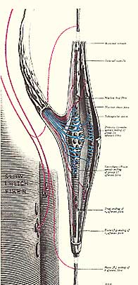
Structure: The spindle is a fusiform-shaped organ, encased in a connective tissue capsule in parallel with a fascicular group of muscles. Each muscle spindle cell is built around 6 to 14 small intrafusal muscle fibers. Like extrafusal muscle fibers, intrafusal muscles are encased in endomysium, and capillaries run between them. |
There are three kinds of intrafusal fibers: bag 1 (dynamic), bag 2 (static), and chain fibers. Only the ends of the intrafusal fibers contract, as the central portions contain no actin or myosin filaments, only nuclei, so the central fibers function as sensory receptors, not contractile units. Little gamma efferent (motor) nerves also feed into the spindle, attaching to both ends of the intrafusal spindle fibers. The alpha motor nerve branch to the extrafusal fibers gives off gamma 1 efferent nerves to the dynamic bag 1 fibers, whereas gamma 2b to the static bag fibers and gamma 2c to the chain fibers come directly from the cerebellum. They all excite mechanical contraction of these intrafusal fibers, just as the alpha motor nerves excite contraction to the extrafusal fibers.
There are also gamma afferent (sensory) nerves leaving the spindle, traveling to either the cerebellum or to the spinal cord through the posterior nerve route. Each of these gamma afferents begin with nerve end organs attached to the intrafusal muscle fibers within the spindle. These end organs are called flower spray and annulospiral end organs.
And lastly, the muscle spindle cell has one lymph duct attached to it, designed to draw fluid, i.e. lactic acid, out of the spindle. Conventional wisdom says that skeletal muscles become inundated with lactic acid after strenuous use, but when muscles stop contracting, and that with enough time, the flow of blood eventually flushes the lactic acid out of the tissues. But we know that not all of the lactic acid gets flushed out; some remains trapped in the spindle cells. Why is this?
The lymph ducts, part of the circulatory system, utilizes skeletal muscles to pump lymph fluid back towards the heart. Once the level of lactic acid has receded in the surrounding muscle tissue, the duct then acts to help flush the spindle and dilute the acid. Nevertheless, in people with hypertonic muscle, during intense activity the levels of acid can accumulate to the point that muscle contraction becomes set in, almost locked, for a period of time. This contraction both continues to produce lactic acid and blocks circulation in the veins; the veins are susceptible because they have such thin walls and so little blood pressure. The lymph duct is actually trying to remove the lactic acid, but when the surrounding muscle is so full of it, the duct ends up drawing it into the spindle. You wouldn’t want to have the air circulator intake on your car, for example, next to the exhaust. So that is primarily why the spindle membrane acts as an accumulating repository for lactic acid.
In doing NeuroSoma®, we know when we are releasing trapped lactic acid because of the almost immediate histamine reaction of redness, whealing, or welts on the skin. The amount of skin irritation depends on two factors: the first is the level of sickness in subject’s suboccipital muscles; very spastic suboccipitals produce large amounts of histamine. And the second is how far away from the surface, how deep into the belly, the release is occurring. The deeper it is, the less likely we are to see the reaction at the surface. So only in the early stages of treatment will we see a very rapid or large surface response. Then as we get more and more layers of soft muscle, we see less and less of it. It’s still happening, but it’s not reaching the surface where we can see it.
Function: The muscle spindle acts as a mechanical synapse. It receives electrical, efferent signals through the gamma efferents which it converts into mechanical muscle contraction. Then the annulospiral nerve end organs take that mechanical reaction – contraction - of the spindle, summates it with the action of the main muscle fibers, and converts it all back into an electrical signal that is fed out into the alpha motor nerve into the main muscle fibers, causing them to contract. The flower spray nerve end organs also summate mechanical contraction, but they feed directly back through gamma afferents to the cerebellum with a report on the amount of contraction taking place in their adjoining skeletal muscle. Synapses ordinarily only feed forward from the CNS, and only allow electrical information to be summed; summations are what synapses are about. However the spindle synapse summates both mechanical and electrical signals.
The spindle cell has four assignments, which are 1) control and maintain muscle tone; 2) activate the dynamic stretch reflex mechanism; 3) maintain muscle contraction against the constant force of gravity (the static stretch reflex mechanism); and 4) control fine motor movements. ‘Static’ is in equilibrium, steady, balanced, constant. ‘Dynamic’ and ‘myotatic’ mean the same thing – constantly changing.
NeuroSoma® focuses predominantly on two of these spindle functions. The stretch reflex mechanism is well known and is acknowledged to be of great significance; even so, it is widely ignored by bodyworkers. This mechanism instantly contracts skeletal muscle tissue as a guarding action when the muscle is stretched past a certain set limit, whether that stretch be active or passive. In other words, the stretch reflex mechanism activates when one overstretches one’s own muscles, or when another overpresses into one’s muscle fibers, regardless of how slow the approach. The NeuroSoma® myotherapist therefore applies a limited pressure not deep enough to activate the stretch-reflex mechanism, and so avoids turning on automatic contraction and irritation of the muscle.
Our primary consideration is maintenance of muscle tone by the spindle. Although this is an essential, key spindle function, it is nevertheless not widely understood. It is only rarely that reference connecting the spindle cell to the maintenance of muscle tone can be found, and usually physiologists who are scientists rather than doctors make the connection. In order to understand muscle malfunction, one must first understand muscle function. Likewise, when the function, or physiology of muscle tonus is comprehended in its entirety, it is a short step to understand the malfunction of muscle tone and how to correct it. NeuroSoma® is the product of this knowledge, and although hundreds of therapies promise to “soften” muscle, this bodywork modality actually does turn hard, hypertonic muscle back into healthy soft tissue by utilizing cutting edge science.
|
|
|
|
|
|
|
© ALL CONTENT ON THIS WEBSITE, INCLUDING IMAGES, HTML, AND TEXT ARE PROTECTED; COPYRIGHT 2006 WELL BEING LLC. ALL RIGHTS RESERVED. REPRODUCTTION IN ANY FORM WITHOUT PERMISSION PROHIBITED.
TERMS OF USE
|
|
|
|
|